Introduction

Understanding the protein content of biological samples is essential in various fields, including biochemistry, molecular biology, and clinical diagnostics. BCA (bicinchoninic acid) tables play a crucial role in accurately quantifying proteins, enabling researchers and clinicians to gain insights into protein expression, regulation, and function. In this comprehensive guide, we delve into the intricacies of BCA tables chemistry, exploring its principles, applications, and practical considerations.
Principles of BCA Chemistry
BCA tables utilize the principles of the bicinchoninic acid (BCA) assay, a colorimetric method for protein quantification. The assay involves the reduction of Cu2+ ions by protein molecules in an alkaline environment, leading to the formation of a colored complex. The intensity of the color formed is directly proportional to the protein concentration, allowing for accurate quantification.
Mechanism:
- Protein-Cu2+ Interaction: Proteins form coordination complexes with Cu2+ ions in an alkaline environment.
- BCA Reduction: BCA reduces the Cu2+ ions to Cu+, forming a purple-blue complex.
- Color Formation: The complex formed between Cu+ and BCA has an absorption maximum at 562 nm, which can be measured using a spectrophotometer.
Applications of BCA Tables Chemistry
BCA tables are widely used in various applications, including:
- Protein Quantification in Biological Samples: BCA tables are used to determine the protein concentration in a wide range of biological samples, such as cell lysates, tissue homogenates, and serum.
- Calibration of Other Protein Assays: BCA tables are used as a reference to calibrate other protein assays, such as the Bradford and Lowry methods.
- Protein Purification and Analysis: BCA tables are employed to monitor protein concentrations during purification procedures and analyze purified protein samples.
Advantages and Disadvantages of BCA Tables Chemistry
Advantages:
- High Sensitivity: BCA tables are highly sensitive and can detect protein concentrations as low as 0.05 mg/mL.
- Wide Dynamic Range: BCA tables have a wide linear dynamic range, allowing for quantification of proteins across a broad concentration spectrum.
- Simple and Rapid: The BCA assay is relatively simple to perform and can be completed within minutes.
Disadvantages:
- Interferences: BCA tables can be affected by certain substances, such as detergents, lipids, and reducing agents, which may interfere with the color formation.
- Low Accuracy at Very Low Concentrations: At very low protein concentrations (<0.5 mg/mL), the accuracy of BCA tables may decrease.
- Not Suitable for All Proteins: BCA tables may not be suitable for quantifying certain proteins, such as glycoproteins or proteins with unusual structures.
Table 1: Composition of BCA Reagents
| Component | Concentration | Function |
|---|---|---|
| Bicinchoninic acid | 4% | Forms a colored complex with Cu+ |
| CuSO4 pentahydrate | 0.5% | Source of Cu2+ ions |
| Sodium carbonate | 2% | Creates an alkaline environment |
| Sodium tartarate | 4% | Stabilizes the colored complex |
Table 2: Performance Characteristics of BCA Tables
| Parameter | Value |
|---|---|
| Sensitivity | 0.05 mg/mL |
| Linearity | 0.05-2 mg/mL |
| Accuracy | ±2% |
| Precision | CV < 5% |
| Interference | Detergents, lipids, reducing agents |
Practical Considerations
- Sample Preparation: Samples should be lysed or homogenized in an appropriate buffer that does not interfere with the BCA assay.
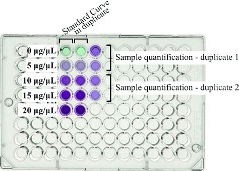
- Standard Curve: A standard curve should be prepared using a known concentration of protein to determine the linear range of the assay.
- Incubation Time: The incubation time for the BCA reaction can vary depending on the manufacturer’s instructions, typically 15-60 minutes at 37°C.
- Color Measurement: The absorbance of the colored complex is measured using a spectrophotometer at 562 nm.
Table 3: Calibration Standards for BCA Tables
| Concentration (mg/mL) | Volume of Standard (μL) |
|---|---|
| 0 | 0 |
| 0.2 | 20 |
| 0.4 | 40 |
| 0.6 | 60 |
| 0.8 | 80 |
| 1.0 | 100 |
| 1.2 | 120 |
Table 4: Troubleshooting Guide for BCA Tables
| Problem | Possible Cause | Solution |
|---|---|---|
| Low Readings | Insufficient incubation time, sample interference | Increase incubation time, use a compatible buffer |
| High Readings | Instrument error, sample contamination | Calibrate spectrophotometer, remove contaminants |
| No Color Formation | Reagent expired, incorrect sample preparation | Replace reagents, prepare samples according to protocol |
FAQs
-
What is the detection limit of BCA tables?
– The detection limit is typically around 0.05 mg/mL. -
Can BCA tables be used to quantify proteins in crude samples?
– Yes, but interfering substances may need to be removed or minimized. -
How stable are BCA solutions?
– BCA reagents are stable for several months when stored at 4°C. -
What factors can affect the accuracy of BCA tables?
– Protein structure, sample preparation, incubation time, and interfering substances. -
Can BCA tables be used to measure protein concentration in cell culture media?
– Yes, but the media should be diluted or supplemented with proteins to minimize interference from nutrients. -
What is the difference between the BCA and Bradford assays?
– The BCA assay is more sensitive and has a wider dynamic range than the Bradford assay. -
What is the future of BCA tables chemistry?
– Ongoing research focuses on improving sensitivity, reducing interference, and developing novel applications. -
What novel applications can be explored using BCA tables?
– Proteomics: Quantifying protein expression profiles in different biological systems.
– Drug Discovery: Identifying targets for drug development by analyzing protein expression changes.
– Diagnostics: Developing biomarkers for disease detection and monitoring.